
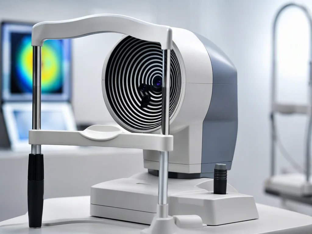
Corneal mapping: understanding shape
Topography and tomography help describe the cornea’s curvature, elevation and structural patterns. They can inform a customised plan and reveal irregularity that makes laser treatment unsuitable.
Reliable capture and clinical interpretation matter as much as the device name. Poor-quality scans may need to be repeated.
Thickness and refraction: planning within limits
Pachymetry measures corneal thickness, while careful refraction establishes the focusing error. Together with the proposed treatment, these measurements help the surgeon consider tissue limits and the correction plan.
No single thickness or power number should be used in isolation to approve surgery.
Ocular surface and retinal assessment
Tear-film and slit-lamp evaluation help identify dry eye and anterior-segment problems. Eye pressure and retinal examination contribute to a broader safety assessment. Additional imaging may be used when clinically indicated.
The exact workup varies between patients and procedures.
No invented machine-model claims
This website does not name manufacturers, model numbers or latest imported labels without current hospital verification. During your consultation, ask which technology will actually be used for your tests and procedure, and why it is appropriate.
Medical information & sources
This page provides general patient education and does not diagnose candidacy or replace an ophthalmologist’s examination. Clinical content requires final treating-doctor approval before launch.